 Precision Machinery Technology Co., Ltd")

Researchers do not struggle because they lack images. They struggle because too many imaging methods force a compromise between visibility and cell health. In many laboratories, the moment a team starts adding dyes, labels, or repeated handling steps, the risk of disturbing the natural condition of the sample rises. That is exactly why interest in the 3D Cell Microscope continues to grow. For teams that want a clearer view of live cells without pushing them too far away from their original state, this approach offers a far more practical path.
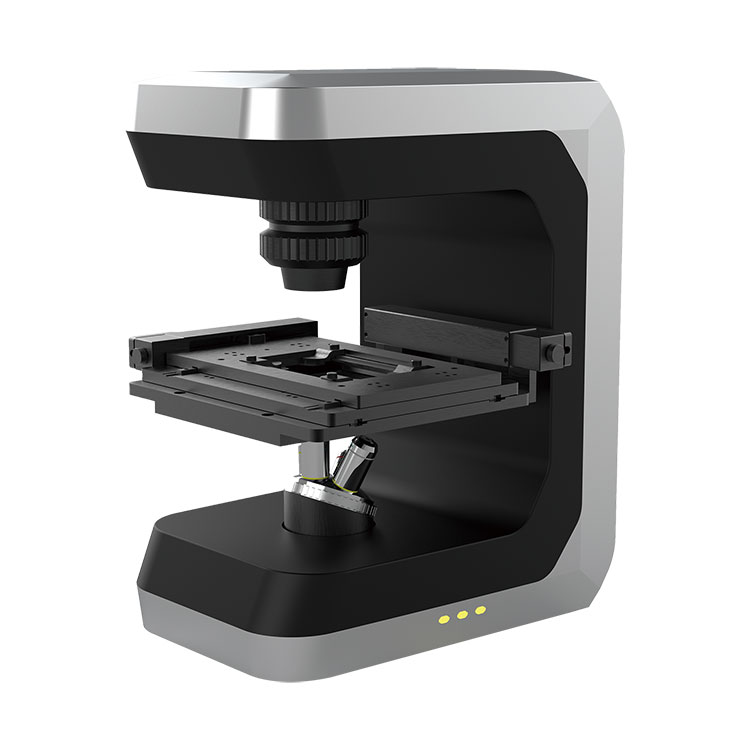
At the center of that conversation is Bojiong (Shanghai) Precision Machinery Technology Co., Ltd, a company focused on precision optical and cell observation solutions. When laboratories need better long-term observation, easier workflow control, and more useful morphology data, a modern 3D Cell Microscope becomes much more than a piece of equipment. It becomes a decision-making tool that supports daily research, reduces avoidable disruption, and helps teams work with greater confidence.
This article explains why a 3D Cell Microscope matters for laboratories that need reliable live-cell observation, reduced sample interference, and stronger image-based analysis. I look at the most common pain points in cell research, compare traditional imaging challenges with the practical value of 3D label-free observation, and show where this type of system can support drug discovery, stem cell work, cell culture monitoring, and morphology-based research. I also include a simple outline, a jump-link table of contents, a comparison table, and a FAQ section to make the article easier to use in real work settings.
Anyone who works with living cells knows the frustration. The more carefully we want to study cell behavior, the easier it becomes to interfere with that behavior. A sample may look fine at the beginning of an experiment, but repeated staining, light exposure, transfers, or environmental changes can gradually alter the condition we were trying to document in the first place.
That creates several familiar problems:
This is where the conversation changes. A laboratory does not only need magnification. It needs a way to observe cells repeatedly, gently, and usefully. That need is what gives the 3D Cell Microscope its real importance.
The practical strength of a 3D Cell Microscope is not just that it can create a three-dimensional view. Its real value is that it supports more natural observation of living samples while still giving researchers meaningful image information. In other words, it helps laboratories stop choosing between “good data” and “gentle handling.”
For many teams, that translates into four direct gains.
That combination is why a 3D Cell Microscope is increasingly appealing to buyers who are tired of workaround-heavy systems. They are not only looking for images. They want a system that fits the reality of modern biological work.
Many buyers already own standard imaging tools, so the real question is not whether traditional microscopes still have value. They do. The question is whether those tools are enough for current live-cell demands. In many cases, they are not enough on their own.
| Observation Factor | Conventional Approaches | 3D Cell Microscope Advantage |
|---|---|---|
| Sample preparation burden | Often requires more preparation steps, especially when contrast or labeling is needed | Can support more direct observation workflows with less sample disturbance |
| Suitability for live cells | Useful, but repeated handling may affect fragile samples | Better aligned with long-term live-cell monitoring needs |
| Depth and morphology insight | May provide limited structural impression in routine observation | Offers richer three-dimensional morphology-related information |
| Long-duration tracking | Possible, but workflow complexity may increase over time | More practical for continuous observation and dynamic change recording |
| Post-observation usability of samples | Can be reduced when dyes or stronger intervention are involved | Supports gentler handling, helping preserve sample value for later study |
What buyers often realize after this comparison is simple: the issue is not whether older methods still work. The issue is whether they work efficiently enough for today’s demands. When a team needs reliable observation of living cells over time, a 3D Cell Microscope often becomes the more sensible next step.
A strong technology earns its place by being useful across real scenarios, not by sounding impressive in theory. That is why buyers usually ask where the system will create measurable value in practice. The answer is broad, but several areas stand out.
These use cases matter because they reflect the same customer pain point from different angles: buyers want to understand living systems without forcing them into an artificial state too early. A 3D Cell Microscope supports that goal in a way that feels practical rather than theoretical.
Not every buyer is looking for the same configuration, so asking the right questions before purchase matters just as much as comparing product names. I usually suggest focusing on the workflow first and the specification sheet second. A system that looks strong on paper may still be a poor fit if it does not match the laboratory routine.
| Buyer Question | Why It Matters |
|---|---|
| Will the system support long-term live-cell observation? | This affects whether the microscope can serve dynamic studies instead of only short imaging tasks. |
| How easy is the workflow for daily operators? | Ease of use reduces training burden, operating variation, and downtime caused by avoidable mistakes. |
| Can the system handle delicate or sensitive cell types? | This is critical for primary cultures, stem cells, and other demanding biological samples. |
| Will the output support both observation and analysis? | Research teams need information they can interpret and compare, not only images that look sharp. |
| Is technical support available when research needs change? | A microscope is not just a purchase. It is part of a longer operational relationship. |
For many buyers, the best decision comes from thinking beyond the initial demonstration. The right 3D Cell Microscope should still make sense after weeks of use, after multiple operators touch it, and after project priorities shift.

Buying the system is only the beginning. A laboratory gets the most value when it builds a clean observation routine around the instrument. Even the best equipment performs below its potential when the workflow remains inconsistent.
I recommend a few practical habits:
When those habits are in place, the 3D Cell Microscope stops being a specialized device that is used only occasionally. It becomes part of the laboratory’s regular thinking process, which is exactly where its commercial and research value grows strongest.
What is the main reason laboratories choose a 3D Cell Microscope?
The most common reason is the need to observe living cells with less disturbance while still gaining useful structural and dynamic information. Laboratories want clearer insight without forcing samples through overly invasive preparation steps.
Is a 3D Cell Microscope only useful for advanced research centers?
No. It is useful anywhere teams need more reliable live-cell observation, especially when traditional workflows are slowing research, affecting sample quality, or limiting long-term tracking.
Can this type of system help with delicate cell samples?
Yes. That is one of its strongest advantages. Laboratories working with sensitive cultures often value a system that supports gentler observation and reduces unnecessary sample stress.
Why does long-term observation matter so much?
Because many meaningful biological changes do not happen in a single moment. Growth, migration, response, and morphology evolve over time. If the imaging method cannot follow that process well, the picture remains incomplete.
How do I know whether a 3D Cell Microscope fits my laboratory?
Start with your daily workflow. If your team needs repeated live-cell observation, gentler handling, clearer morphology insight, and stronger consistency across experiments, the fit is usually very strong.
If your team is still balancing image quality against cell condition, it may be time to rethink the tools behind your workflow. A well-chosen 3D Cell Microscope can help reduce preparation burden, improve observation continuity, and make your cell research more dependable from day to day.
Bojiong (Shanghai) Precision Machinery Technology Co., Ltd is ready to support laboratories that want a more practical and research-friendly way to study living cells. If you are comparing systems, planning a lab upgrade, or looking for a better solution for long-term live-cell observation, contact us today and let us help you find the right direction for your application.

 900-1200nm")


