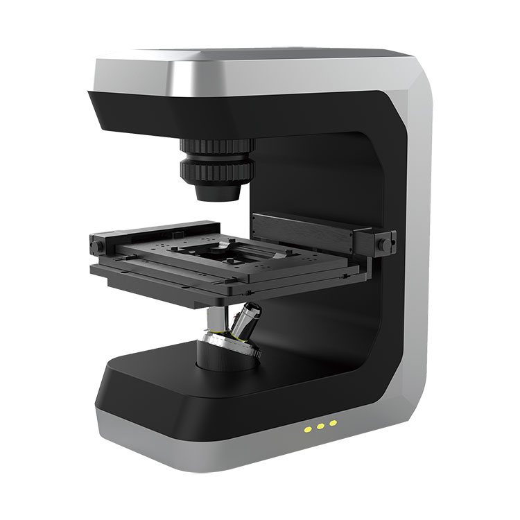
With years of experience in production 3D Live Cell Imaging Microscope, BOJIONG can supply a wide range of 3D Live Cell Imaging Microscope. High quality 3D Live Cell Imaging Microscope can meet many applications, if you need, please get our online timely service about it. In addition to the product list below, you can also customize your own unique 3D Live Cell Imaging Microscope according to your specific needs.We have a professional team composed of scientists from Zhejiang University and Singapore Nanyang Technological University, which provides a strong guarantee for 3D Live Cell Imaging Microscope technology application and innovation.
Advantages of 3D Live Cell Imaging Microscope:
No damage - no labeling or staining, no light toxicity, reducing the true state of cell growth.
Small size - long-term stable operation in the incubator, and time-lapse imaging analysis under normal physiological conditions.
Real-time - Monitoring individual cells in real time while obtaining population data and providing complete quantification of cell morphological parameters.
Multiple data - The same sample or result can be reanalyzed to obtain more experimental data.
Widely used - can be used for cell proliferation, differentiation, migration, apoptosis, toxicology and other aspects of research.
Intelligent - Simple and practical intelligent image analysis software, automatic high-content quantitative analysis of the obtained images.
3D Live Cell Imaging Microscope is a new product for biological and medical cell culture research and detection. It can make up for the shortcomings of phase contrast microscope, fluorescence microscope and confocal microscope in 3D quantitative phase, in vivo and high efficiency, realize cell biopsy, cell count, morbid screening of red blood cells, imaging of cancer living tissue, and provide quantitative analysis of 3D morphology and quality of cell contour information. Applications: Cancer research, stem cell research, drug discovery and development, and in vitro wound healing validation.


