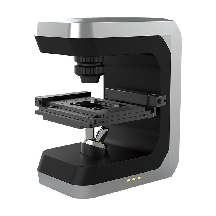
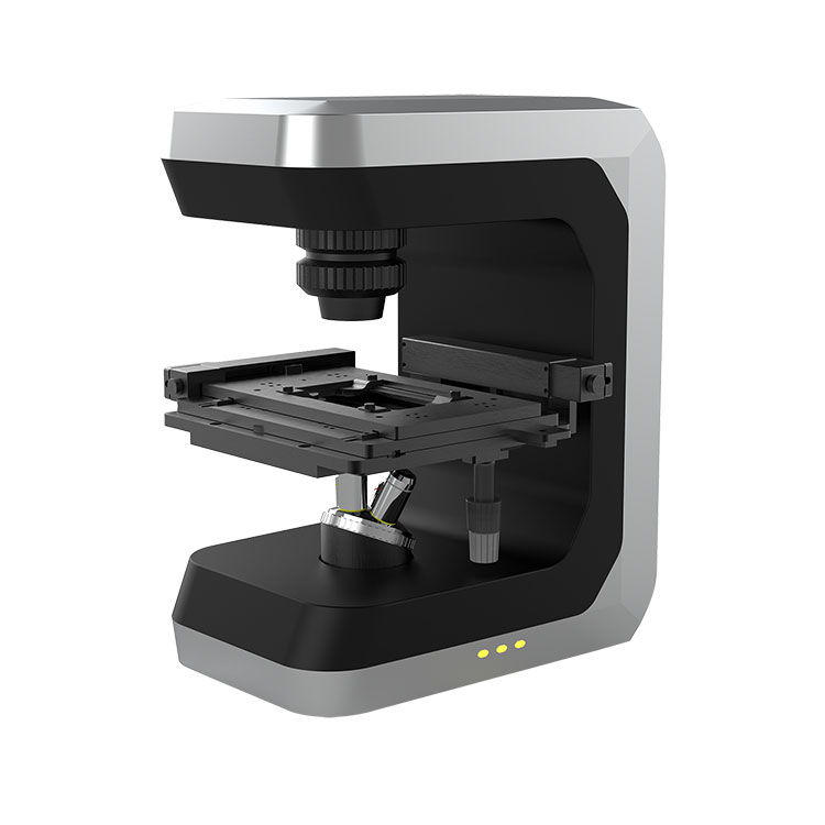
BOJIONG is committed to the production and sales of 3D Living Cell Microscope, with strong technical support, excellent quality and service guarantee. Support product keyword customization! We have our own factory, with a professional team composed of scientists from Zhejiang University and Singapore Nanyang Technological University, with nearly 30 years of rich experience and profound theoretical foundation in the field of interference detection, which provides a strong guarantee for the technical application and innovation of 3D Living Cell Microscope.Welcome new and old customers to continue to cooperate with us to create a better future!
3D Living Cell Microscope adopts phase interference imaging technology, using natural light as coherent light source, using four-wave interference principle to record the phase and amplitude information of the sample to complete the imaging of cells. Therefore, there is no need to process cells, reduce human intervention, and obtain quantitative parameters and high-quality images and data at both single cell and population levels without labeling and invasion. Interference detection with miniaturization and high vibration resistance is realized, which is especially suitable for high-precision detection in biological, industrial, scientific research and other fields.
3D Living Cell Microscope provides a fast, accurate and non-invasive solution for the automatic counting of adherent cells in incubators. Graphically present images and results in real time, generating growth curves over time while monitoring cell morphology, proliferation rate, and confluence. It is widely used in the study of living cell proliferation, the detection of living cell motility and the observation of living cell dynamic morphology. Application in cancer research: It can observe the behavior changes of cancer cells in different environments in real time and evaluate the effect of anti-cancer drugs. Application in stem cell research: The differentiation process of stem cells under different culture conditions was observed through 3D Living Cell Microscope, and the development mechanism of stem cells was studied. Application in drug discovery and development: 3D Living Cell Microscope is used to observe the direct effects of drugs on cancer cells or infected cells, so as to quickly screen potential therapeutic drugs.


