 Precision Machinery Technology Co., Ltd")

3D cell microscopes have revolutionized biological and medical research by providing unprecedented insights into cellular structures and processes. These advanced imaging systems allow scientists to observe cells in their native, three-dimensional state, leading to more accurate and meaningful data. Below, we explore the core application areas where 3D cell microscopes are making a significant impact.
Cancer Research
3D cell microscopes enable detailed analysis of tumor spheroids and cancer cell behavior in a microenvironment that closely mimics in vivo conditions. This helps in understanding metastasis, drug resistance, and treatment efficacy.
Neuroscience
Researchers use these microscopes to study neural networks, brain tissue, and neuronal development in 3D, providing insights into neurodegenerative diseases like Alzheimer’s and Parkinson’s.
Stem Cell Research
Observing stem cell differentiation and organoid development in three dimensions is critical for regenerative medicine and developmental biology. 3D cell microscopes offer the precision needed to track these complex processes.
Drug Discovery and Toxicology
Pharmaceutical companies rely on 3D cell imaging to screen drug candidates and assess their effects on 3D cell models, reducing the need for animal testing and improving prediction accuracy.
Immunology
Studying immune cell interactions, such as T-cell responses and antigen recognition, in a 3D context provides a deeper understanding of immune system mechanisms and vaccine development.
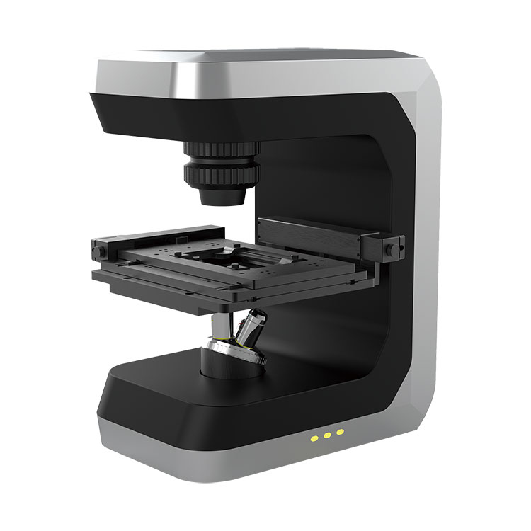
Our 3D cell microscopes are engineered for high performance, reliability, and versatility. Here are the key parameters that define our cutting-edge systems:
| Feature | Specification |
|---|---|
| Resolution | Up to 140 nm laterally, 300 nm axially |
| Imaging Modality | Confocal, Light Sheet, or Super-Resolution options available |
| Sample Compatibility | Supports live cells, fixed samples, and organoids up to 2 mm in size |
| Objective Lenses | High NA objectives (e.g., 40x/1.3 NA, 60x/1.4 NA) for superior clarity |
| Camera System | sCMOS camera with high quantum efficiency and low noise |
| Software | User-friendly interface with AI-assisted analysis and 3D reconstruction capabilities |
| Environmental Control | Temperature, CO₂, and humidity regulation for long-term live-cell imaging |
| Throughput | Automated multi-well plate imaging for high-content screening |
Superior Imaging Quality: Capture detailed 3D structures with minimal phototoxicity.
Flexibility: Adaptable to various imaging modes and sample types.
Efficiency: Streamlined workflows reduce time from sample preparation to data analysis.
In summary, 3D cell microscopes are indispensable tools across multiple research domains, from cancer biology to immunology. Their ability to deliver high-resolution, lifelike imaging transforms how scientists study cellular mechanisms. With robust features and adaptable designs, our 3D cell microscopes meet the highest standards of modern laboratories, driving discoveries that were once beyond reach.
Invest in our 3D cell microscopes to elevate your research capabilities and stay at the forefront of scientific innovation. If you are very interested in Bojiong (Shanghai)Precision Machinery Technology's products or have any questions, please feel free to contact us.


")


